THE BRAIN, IN PRACTICE
Learning neuroscience through observation, dissection, and experiment.
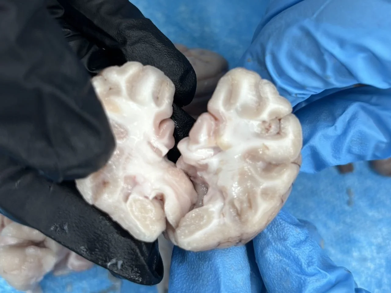
I explored the nervous system through hands-on experiments, from simulated cataract surgery and pig-brain dissection to investigating phototaxis in Hydra and conducting reflex experiments with spinal frogs. I also used RT-PCR to compare gene expression patterns between pig muscle tissue and brain tissue, examining how molecular differences reflect their distinct biological functions.
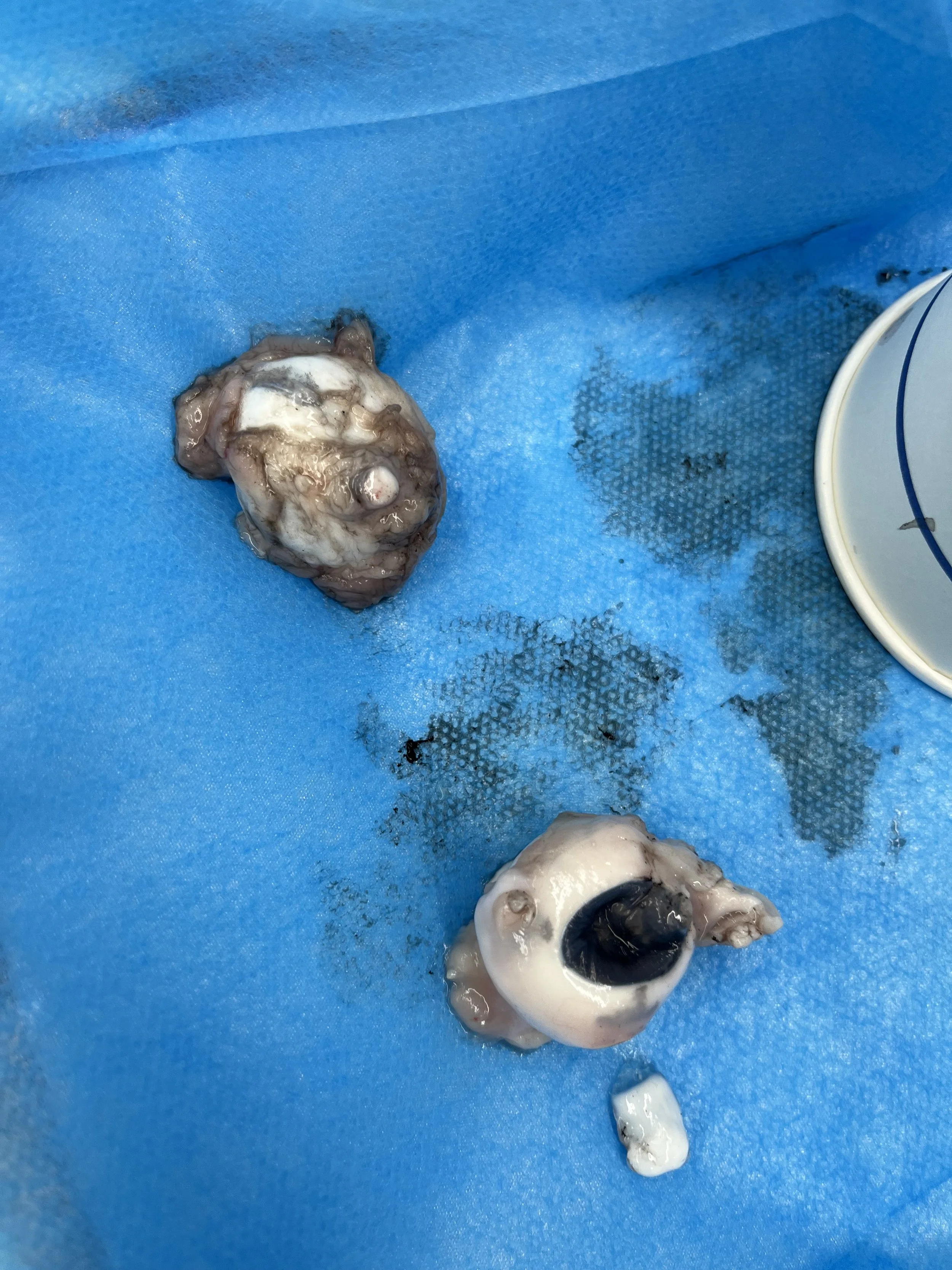
Simulated Cataract Surgery
Using a pig eye as an anatomical model, I explored the basic principles and workflow of cataract surgery through a hands-on simulation.
01 — Inducing Cataract-like Opacity
The pig eye was briefly heated in a microwave for approximately 10 seconds to create a visible lens opacity, simulating the clouding characteristic of a cataract.
02 — Corneal Incision
I made an incision through the cornea to gain access to the lens, learning how the eye’s anterior structures provide a pathway for surgical intervention.
03 — Lens Replacement
After accessing the lens, I removed the simulated cloudy lens and replaced it with a clear artificial lens, modeling the central objective of cataract surgery: restoring the optical pathway.
04 — Closure
The incision was carefully closed with sutures, completing the simulated procedure and reinforcing the importance of precision and tissue handling in ophthalmic surgery.
RT-PCR: From Tissue to Molecular Identity
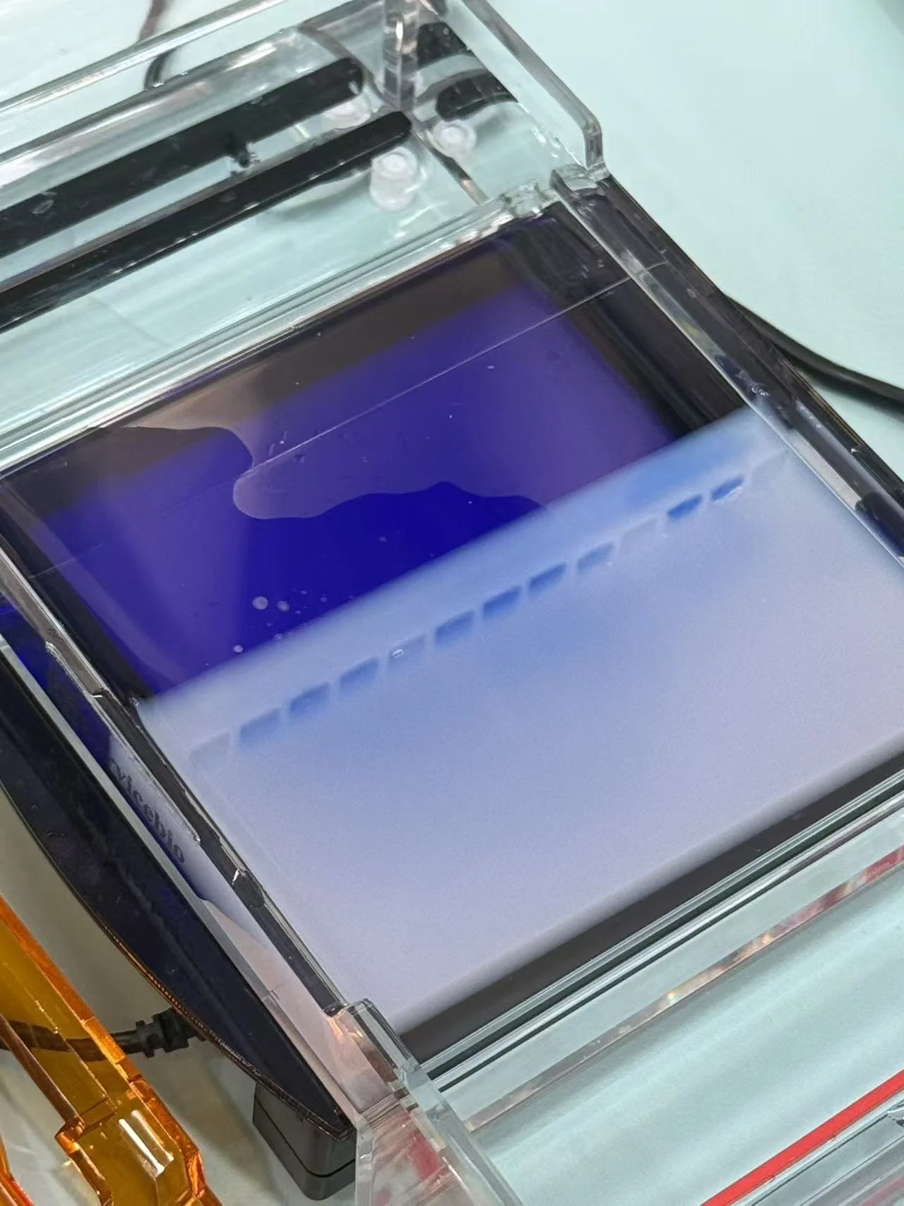
To compare pig brain and muscle tissue at the molecular level, I used RT-PCR to examine differences in gene-expression patterns. After extracting RNA and converting it into cDNA, I analyzed neural- and tissue-associated markers including MBP, MT3, and LSAMP, with HK1 and GAPDH as reference genes. Gel electrophoresis allowed me to visualize the amplified products and connect molecular expression patterns with the distinct biological functions of brain and muscle tissue.